
 Kontakt
 Kako kupovati

Dostava
Savjetnik za kupnju
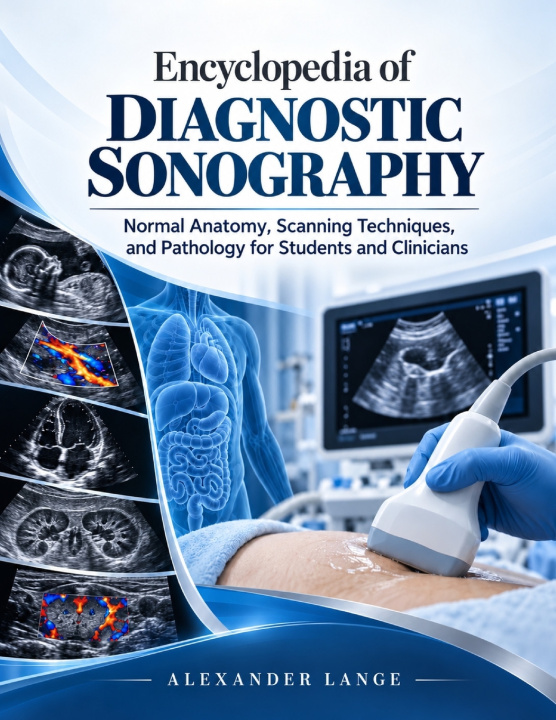
Encyclopedia Of Diagnostic Sonography
Normal Anatomy, Scanning Techniques, and Pathology for Students and Clinicians
 Engleski
 317 b
 Novo
Do 30 dana za povrat

Ultrasound becomes challenging when anatomy, probe position, image optimization, Doppler, and pathology must all be interpreted at the same time. A structure may look different because of angle, gain, patient habitus, artifact, normal variation, or disease-and learning to distinguish among those possibilities is central to confident scanning and sound clinical reasoning.
This comprehensive reference connects ultrasound physics with the practical workflow of the examination. It begins with acoustic principles, transducers, beam behavior, image controls, Doppler fundamentals, artifacts, safety, infection prevention, and ergonomics, then applies those foundations across major organ systems and clinical settings.
Inside, readers will learn how to:
• Select appropriate transducers, patient positions, acoustic windows, and scanning planes.
• Recognize normal sonographic anatomy, expected measurements, and important variants before judging abnormality.
• Optimize grayscale, color, power, and spectral Doppler for the structure and clinical question.
• Evaluate abdominal, hepatobiliary, pancreatic, renal, pelvic, obstetric, vascular, thyroid, breast, scrotal, musculoskeletal, pediatric, neonatal, and emergency examinations.
• Compare common disease patterns, differential considerations, artifacts, pitfalls, and limitations of ultrasound.
• Strengthen documentation, reporting, urgent communication, quality assurance, and understanding of image-guided procedures.
Practical protocols, tables, clinical pearls, caution points, troubleshooting guidance, review questions, and detailed illustrations reinforce the connection between technique and interpretation.
Designed for medical imaging learners and practitioners who need a broad clinical reference, the book supports study, clinical preparation, protocol review, and continued learning. It helps readers connect what appears on the screen with how the image was acquired, what normal should look like, and which findings deserve closer evaluation.
Build a more organized understanding of ultrasound from first principles through advanced clinical applications, with anatomy, technique, Doppler, pathology, and clinical workflow brought together in one substantial resource.


Informacije o knjizi
Engleski

Poklonite ovu knjigu još danas
To je jednostavno
1 Dodajte knjigu u košaricu i odaberite isporuku kao poklon 2 Zauzvrat ćemo vam poslati kupon 3 Knjiga dolazi na adresu poklonoprimcaDobar dan! Ja sam Libroamiko, vaš književni savjetnik.
Kako vam mogu pomoći?