
 Kontakt
 Kako kupovati
Dostava
Savjetnik za kupnju
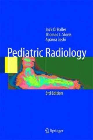
Pediatric Radiology
 Engleski
 244 b
244 b
Do 30 dana za povrat
Kupci su kupili i








a b Fig. 3.50. A child with acute onset of respiratory distress.(See Appendix 2) a b Fig. 3.51. Chronic lung disease.(See Appendix 2) way to read systematically is to evaluate the tech- and diaphragm). Now evaluate Figs.3.50-3.52.- nical factors of lung volume, patient position, and proach each one systematically and describe the - the way the ?lm was exposed. Use the radiologist's normalities you see. The answers are in the App- circle and ABCS: A=abdomen, B=bones, S=soft dix 2. tissue, and C=chest (airway, mediastinum, lungs, 03_Haller_Chest 22.10.2004 13:06 Uhr Seite 62 62 3 Chest Examinations in Children a b c d Fig. 3.52. A child with wheezing.(See Appendix 2) 3. Webb WR, Mueller NL, Naidich DP (1996) High-resolu- References and Further Reading tion CT of the lung,2nd edn.Lippincott-Raven,New York 4. Kuhn JP,Slovis TL,Haller JO (eds) (2003) Caffey's pedi- 1. Slovis TL, Haller JO, Berdon WE, Baker DH, Joseph PM atric diagnostic imaging,10th edn.Mosby,Philadelphia (1979) Noninvasive visualization of the pediatric airway.


Informacije o knjizi
Engleski

Poklonite ovu knjigu još danas
To je jednostavno
1 Dodajte knjigu u košaricu i odaberite isporuku kao poklon 2 Zauzvrat ćemo vam poslati kupon 3 Knjiga dolazi na adresu poklonoprimcaMoglo bi vas zanimati i

 /
/
 Meki uvez
 43.33
€
43.33
€


 /
Meki uvez
38.19
€
/
Meki uvez
38.19
€









Dobar dan! Ja sam Libroamiko, vaš književni savjetnik.
Kako vam mogu pomoći?